What is magnetic resonance imaging?
Magnetic resonance imaging (MRI/MRI) is an imaging procedure that works with strong magnetic fields and radio waves. In contrast to computed tomography (CT), it does not involve any radiation exposure. MRI is also known as magnetic resonance imaging. Similar to CT, radiologists create detailed, three-dimensional cross-sectional images of the inside of the body. The method is painless.
MRI makes use of the fact that water molecules (hydrogen to be precise) in the human body arrange themselves in parallel under a strong magnet. When radio waves hit it, the orientation of the hydrogen nuclei changes. After the radio wave impulse, they return to their parallel starting position. The extent of this deflection can be recorded and converted into layer images by a computer.
We can diagnose diseases and injuries to organs, soft tissue and tissue particularly well using MRI. However, hard structures such as bones or joints can also be visualized – albeit less precisely because they contain fewer water molecules. Depending on the water content, the structures and tissues appear lighter or darker on the MRI images.
Radiologists usually work with a contrast agent containing gadolinium, which they inject into the vein. This enables them to differentiate between the various tissues even better, for example in the case of tumors, inflammation, signs of wear and tear, injuries or vessels.
How does an MRI work?
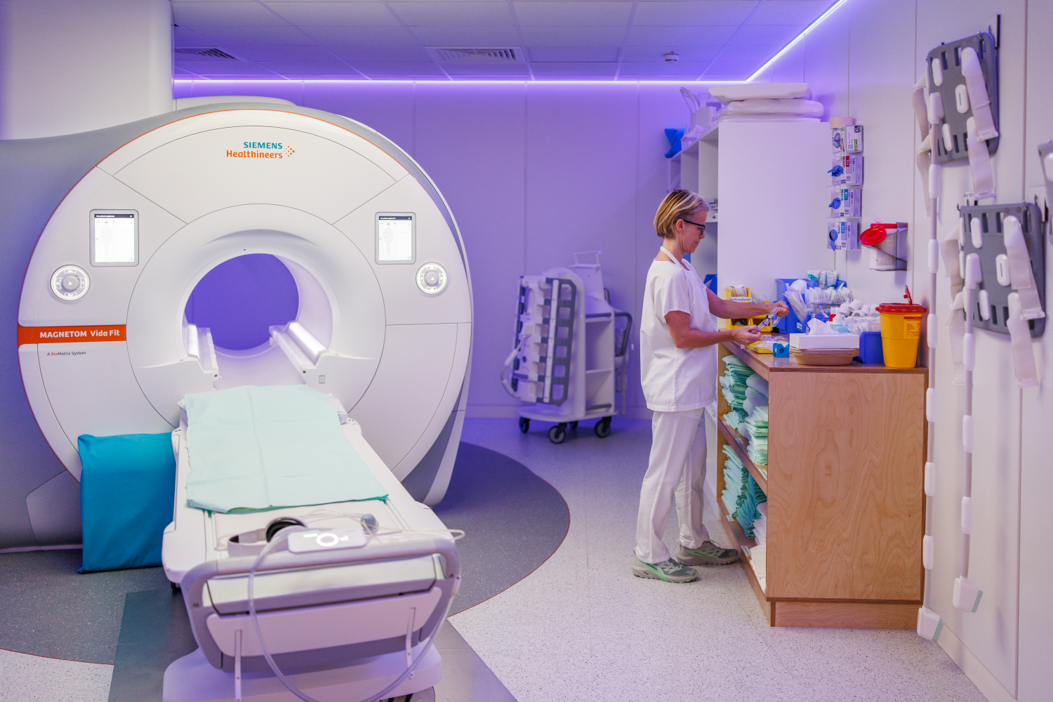
MRI uses a magnetic resonance tomograph – a device that looks like a tube and is narrower than a computer tomograph. People with claustrophobia can be given a sedative in advance.
Electric coils are integrated into the wall of the tube, which generate strong magnetic fields and radio waves. Water molecules (or the hydrogen) in the human body react to the magnetic fields and can be deflected from their path. This produces certain signals, which in turn depend on the nature of the tissue. An MRI scanner measures these signals and sends them to a computer, which calculates three-dimensional sectional images in black and white.
The human body consists of more than 70 percent water. This is stored in different quantities in the organs, tissues and structures: the looser a tissue is, the more water or hydrogen it contains. And: the more water an organ contains, the easier it is to recognize on the MRI image. Because bones contain less hydrogen, they are less visible in the images. There are therefore more meaningful diagnostic methods for bone fractures.
Magnetic resonance imaging - the procedure
In most cases, you do not need to prepare specially for magnetic resonance imaging. In rare cases, you may need to be sober for the examination. If preparation is necessary, you will be informed in advance. Depending on the medical issue, the examination takes about 20 to 30 minutes, sometimes up to an hour.
It is important that you remove or discard all metal items before the examination, such as watches, earrings, piercings, belts with metal buckles or hearing aids. The strong magnet of the magnetic resonance tomograph can attract the metal, cause it to slip or heat it up. This can cause injuries, for example burns. In addition, metallic objects can falsify the test results.
Some metal parts cannot be removed, such as pacemakers, implants, stents in the coronary arteries or contraceptive coils. In this case, an MRI may not be possible. Tattoos, some cosmetics and permanent make-up can also contain traces of metals. Always check with your doctor beforehand whether magnetic resonance imaging is possible.
Some tips for MRI:
- Some patients find it helpful to relax during the examination and imagine a pleasant scene – for example, taking a mental walk in the forest or by the sea.
- Abdominal breathing can also alleviate anxiety and relax some people.
- If you have claustrophobia, you can take a sedative before the examination. You will then notice less of the examination and survive it better.
The MRI procedure can be described as follows:
- You lie down as comfortably as possible on a lounger. The radiology technician will help you and place a so-called receiving coil, which is necessary for image acquisition, on the body region to be examined. You will then be pushed onto the couch into the MRI scanner.
- During the examination you will be alone, but connected to the staff in the next room via an intercom system. You can communicate with the radiology assistants at any time.
- You will be given an emergency button. It offers safety, especially for those with claustrophobia in the narrow tube. If you press the button, you can cancel the examination at any time – however, you will then have to start the MRI again from the beginning.
- You will be given headphones to wear over your ears and/or earplugs for soundproofing, as the examination is quite loud. The appliance produces loud pounding and knocking noises. With a little imagination, the sounds can also be interpreted as music.
- During the examination you must lie as still as possible and must not move.
- Sometimes we inject a contrast agent with gadolinium (a metal) into the vein to increase the contrast of the tissue.
Some hospitals and doctors’ surgeries now work with open MRI machines. The coil is divided into two thick slices between which you lie (as in a “sandwich”). Advantage: You have a clear view of the outside. Disadvantage: The examination often takes longer and the image quality is poorer.
There are also special devices that enable MRI examinations to be carried out in a standing or sitting position, for example for the joints or spine. They are called upright MRI scanners and are also suitable for people who are very overweight. A tube is often too narrow for them.
Areas of application: When is magnetic resonance imaging used?
MRI is used in the diagnosis of various diseases and injuries. Most organs, tissues and soft tissues can be visualized well. Hard structures such as bones and joints, on the other hand, are less easily recognizable due to the low hydrogen content.
Some examples:
- Brain, e.g. stroke, brain tumors, vascular malformations, inflammation, injuries after accidents
- Joint diseases, disc wear and tear, vertebral body fractures
- Diseases of the liver, bile ducts and pancreas
- Cancers, e.g. breast cancer, prostate cancer, colon cancer, liver tumors, lymphomas – We can determine the location, size and spread of the tumor, and also detect metastases in other organs and lymph nodes.
- Diseases of the nerves (MR neurography)
- Inflammation of the soft tissues, in various organs and vessels
- Vascular changes in arteries and veins, e.g. constrictions or occlusions
- Heart diseases
Magnetic resonance imaging can also be combined with other examination methods to obtain even more meaningful results. One example is positron emission tomography (PET-MRI, e.g. for cancer). With the help of MRI, we can also take specific tissue samples (MRI-assisted biopsy, e.g. of the breast or prostate).
MRI: advantages and risks
MRI has several advantages over other examination methods.
The most important are:
- Like ultrasound, it does not involve exposure to radiation.
- The MRI does not cause any pain.
- MRI can be used to diagnose almost all diseases, injuries and changes in organs and tissues.
- The method is particularly suitable for the visualization of soft tissues such as muscles, tissue, etc.
- Magnetic resonance imaging can also be performed on pregnant women. However, you should always discuss with your doctor whether an MRI is really necessary.
MRI also has some disadvantages and risks:
- It is relatively expensive.
- An MRI may not be possible for metal implants that are firmly seated in the body.
- For people with claustrophobia, the closed tube is often very frightening.
- It takes longer than an ultrasound or CT scan, for example.
- Contrast media containing gadolinium can sometimes cause side effects, such as a temporary feeling of warmth or cold, headaches, discomfort, tingling or skin irritation. Allergic reactions to this contrast medium are rare. Repeated administration of contrast media containing gadolinium can lead to deposits of gadolinium in the brain. For this reason, the European Medicines Agency (EMA) has banned certain (linear) contrast agents containing gadolinium or only permits them in exceptional cases. Three linear gadolinium preparations are still approved in Switzerland, but radiologists rarely use them. So-called macrocyclic contrast agents are still permitted. The gadolinium is said to be better bound in them than in the linear agents. However, gadolinium residues can also be detected in the brain after their use. However, it is not certain that they cause long-term damage to health. We always use contrast media in the smallest possible quantities and only if it is necessary to answer the medical question.
- Contrast media containing gadolinium can lead to so-called nephrogenic systemic fibrosis (NSF) – a skin disease – if kidney function is severely impaired. In the case of severely impaired kidney function, contrast media containing gadolinium are only used if necessary to answer the medical question and in the smallest possible quantity.