Diagnosis
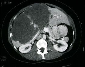
The diagnosis can be made on the basis of characteristic features using ultrasound, CT or MRI. In a few cases, atypical hemangiomas can only be diagnosed by a combination of different imaging techniques.

Computer tomogram of a giant hemangioma of the liver
Treatment
Asymptomatic patients, especially those with lesions < 5 cm, do not require therapy and are observed. Long-term observations have shown that in most cases neither growth nor complications occur. On the other hand, lesions > 5 cm are characterized by rapid growth, which necessitates close radiological monitoring. In asymptomatic patients, the risk of bleeding is too low to justify prophylactic resection.
The treatment of symptomatic hemangiomas or those with a size of > 10 cm requires resection. Various surgical procedures are available, such as enucleation, liver resection or even liver transplantation – the most common procedure is liver resection with preservation of the surrounding liver tissue.

Certificate Publications


