Bleeding in the area of the unstable pelvic ring fractures is often difficult to control. The bleeding can be so extensive that the patient’s life is acutely endangered. In these cases, immediate stabilization of the pelvis is necessary. The urinary organs are also at risk due to the neighboring location. Older patients often suffer so-called stable fractures (over 50% of pelvic fractures) as a result of falls (low-energy trauma).
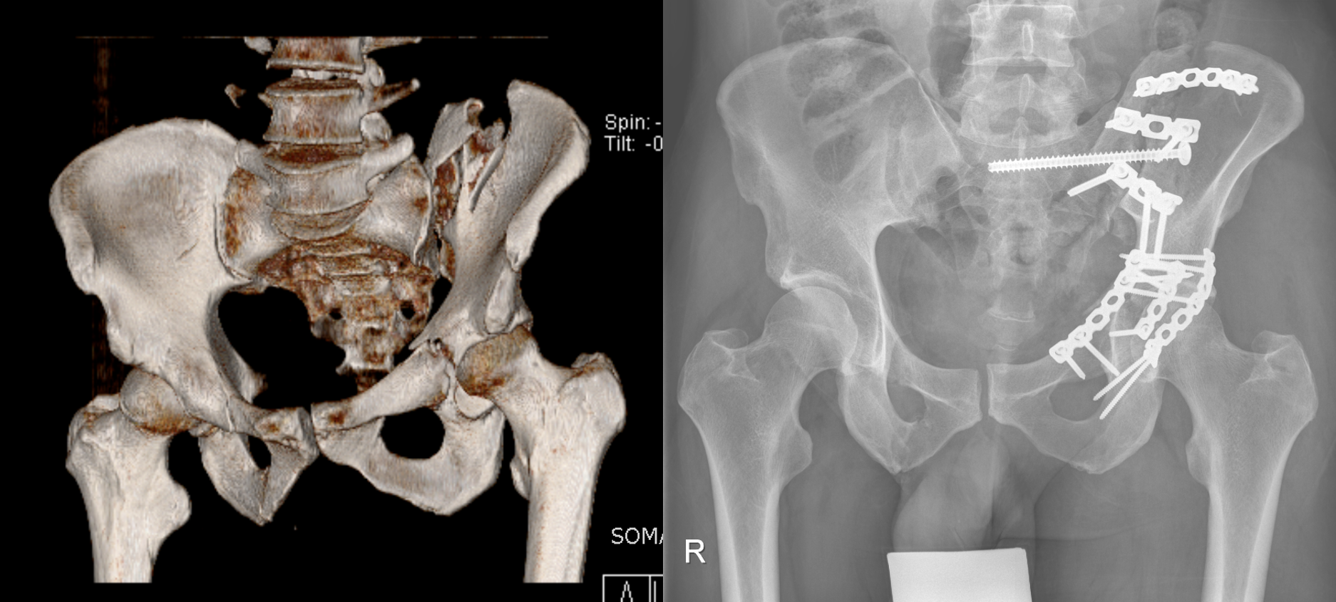
Left: 3D image of a CT scan after combined left-sided pelvic and acetabular fracture. Right: Postoperative X-ray after fixation with plates and screws.
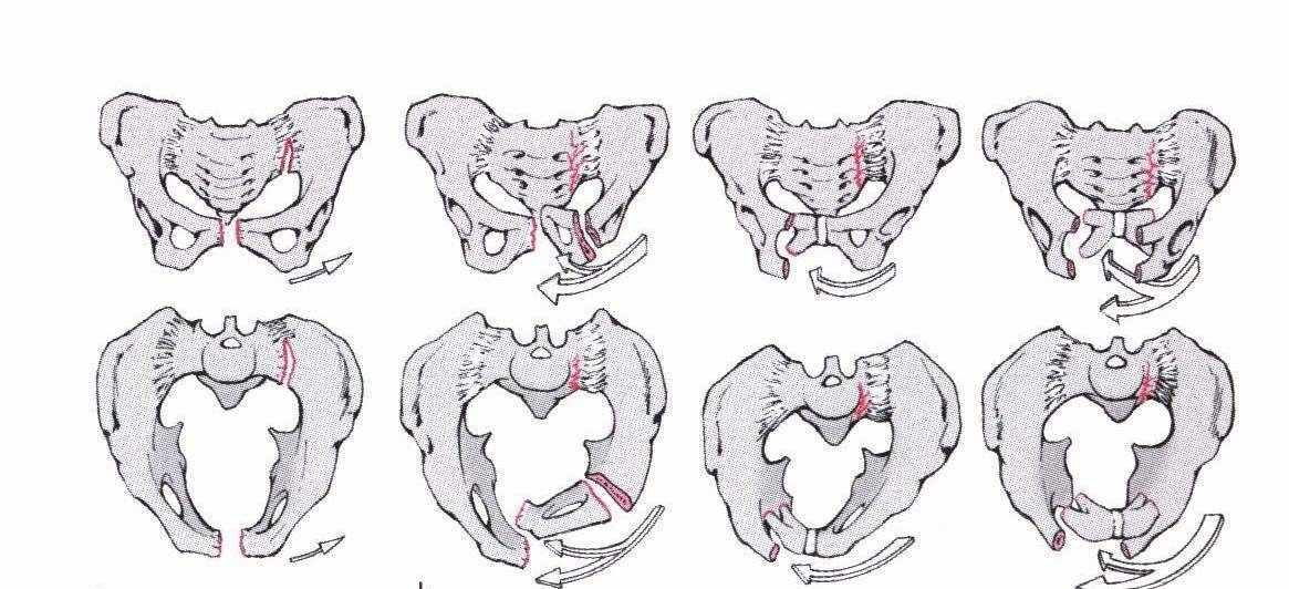
The underlying forces are divided into
- Compression from the side
- Compression from front to back
- Protection in the vertical direction (along the spinal column axis)
- Combination injuries
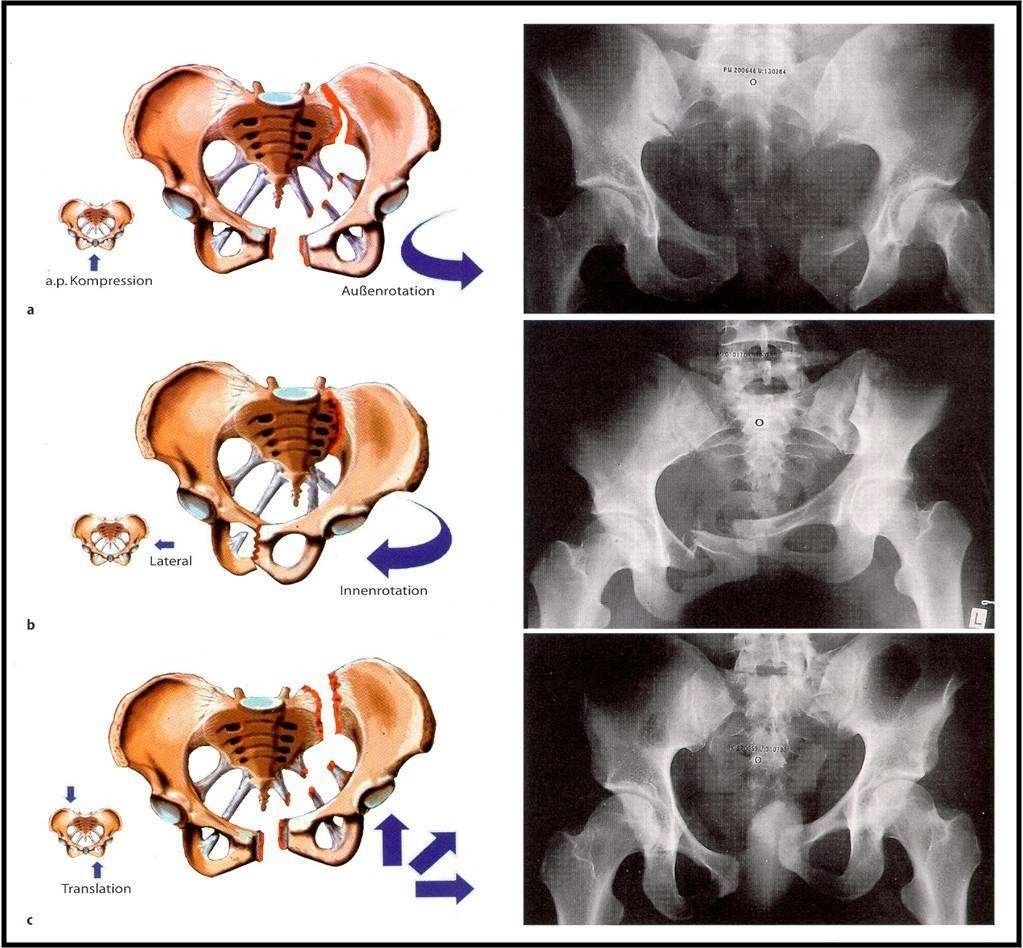
Acting forces and resulting fractures (Orthopaedics and Trauma Surgery up2date, Thieme)
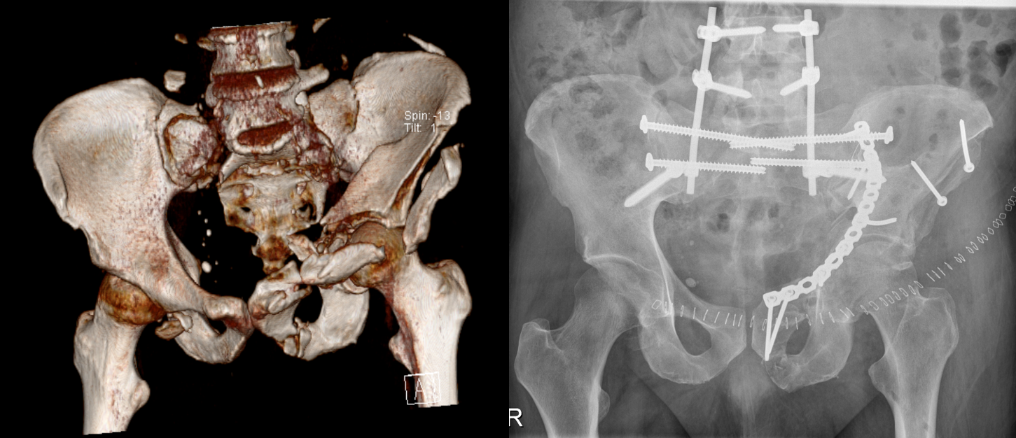
Left: 3D image of a CT scan after a very severe bilateral combined pelvic and acetabular fracture with separation of the pelvis from the lumbar spine. Right: Postoperative X-ray after restoration of the alignment of the hip and pelvic ring and the connection to the lumbar spine using plates, screws and rods.
A basic distinction must be made between stable and unstable pelvic fractures. Important:
- The pelvis is stable when the essential structures of the posterior pelvic ring are uninjured.
- The pelvis is unstable if bony or ligamentous structures in the area of the anterior and posterior pelvic ring are injured.
There are various classification systems, but ultimately they all differentiate between stable, rotationally unstable and rotationally/vertically unstable.
Stable injuries are fractures or avulsions of the ilium, ischium, pubis or coccyx below the joint connections that do not jeopardize the stability of the pelvic ring. These injuries can often be treated without surgery. The focus here is on combating pain and mobilizing the patient.
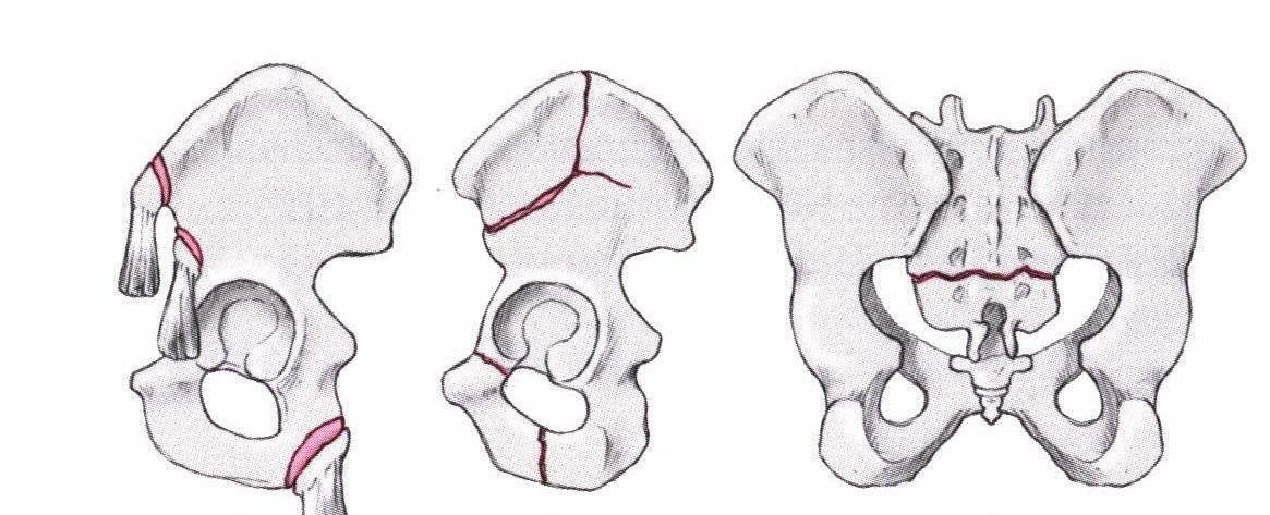
Stable pelvic fractures (Ficklscherer, Orthopaedics and Traumatology, Urban & Fischer)
Rotational instability occurs, for example, when the anterior parts of the pelvis are disrupted or the posterior structures are displaced. Here, however, the vertical stability is maintained. An externally rotated half of the pelvis is referred to as an open-book injury (see Figure 5). These fractures are treated conservatively or surgically, depending on the extent of the injury.
Rotationally unstable fractures (Ficklscherer. Orthopaedics and Traumatology, Urban & Fischer)
Open-book violation
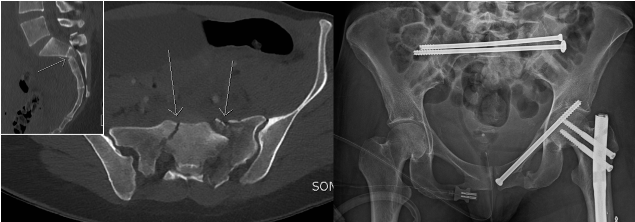
Left: Computer tomography after break-out injury of the first sacral vertebra. Right: Postoperative X-ray after minimally invasive stabilization using long screws through small skin incisions.