Especially in the early diagnosis of sclerodermaan autoimmune multisystemic disease, it has an outstanding role, as disorders of the capillary architecture can often precede other signs of the disease by decades. However, it is also a helpful method for diagnosing other diseases from the group of collagenoses group. For example, very typical images can also be shown for mixed collagenosis or dermatomyositis.
Capillary microscopy can also be used to assess the severity of the circulatory disorder by evaluating the number of capillaries and based on signs of dysangiogenesis, thus estimating the risk of ulcers occurring.
How is the examination carried out?
The method is completely painless and non-invasive. The smallest blood vessels (capillaries) lie just below the surface of the nail bed and are moistened with highly pure oil to make them more visible.
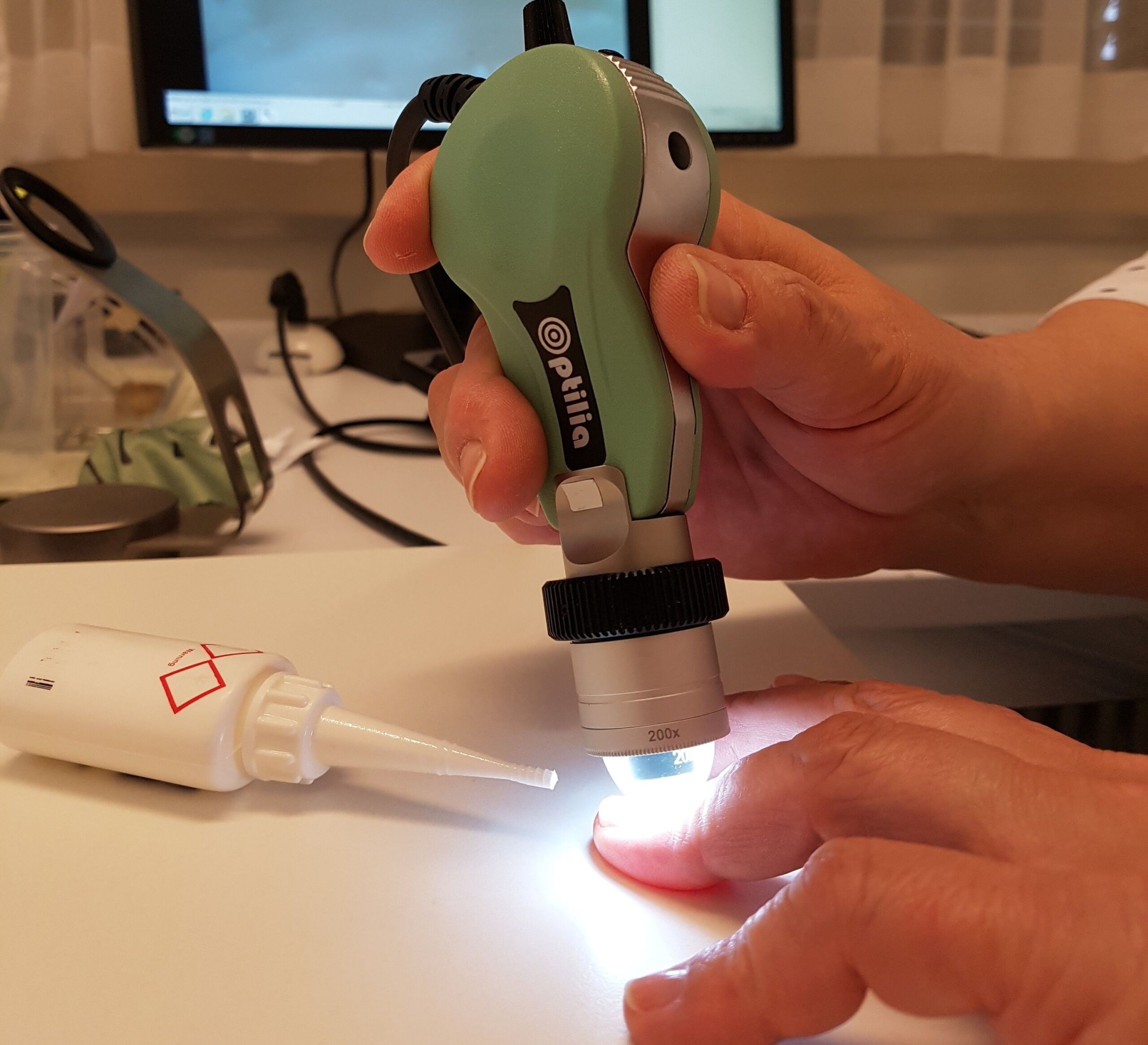 Modern video capillary microscopy is used to examine the blood flow and condition of the capillaries and to count, measure and assess them according to their shape.
For which diseases should a capillary microscopy be performed?
A typical indication for microscopy of the capillaries is the clarification of Raynaud’s syndrome (whitening, blueness, redness of the fingers when exposed to cold or nervousness), which is very often harmless as it is purely functional, but can sometimes be the first sign of collagenosis. This is known as a secondary Raynaud’s phenomenon, which is very common in certain collagenoses.
Patients for whom no further investigations are required can be specially referred for capillary microscopy.
Capillary microscopy is carried out at the Clinic for Rheumatology both on campus and at the Circle (airport).