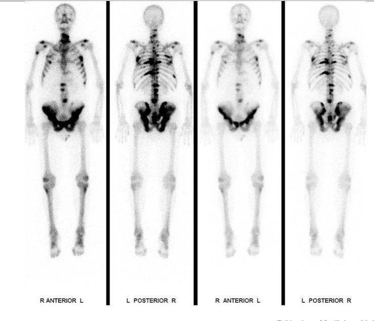
Skeletal scintigraphy for multiple bone metastases
Skeletal scintigraphy is used to search for skeletal metastases, rheumatologic diseases, inflammations and infections as well as suspected child abuse.

Skeletal scintigraphy for multiple bone metastases
At the beginning of the examination, a slightly radioactive substance is injected into a vein in your arm. This substance accumulates in your skeleton and we can use it to examine your bone metabolism.
The images are taken approx. 3 hours after the injection (takes approx. 30 – 60 minutes). In the meantime, you can leave the institute. You may eat normally and should drink at least 1-2 liters of water (no dairy products) in the meantime.
In certain cases, we also examine the soft tissue perfusion by recording a so-called early phase. These early images are taken immediately after the injection or during the injection and last 30 minutes.
The radiation exposure of the examination is comparable to the annual, natural radiation exposure (4 mSv) and is not increased by the number of images. If you are pregnant or may be pregnant, or if you do not know for sure, please report this before the examination. Please also note that you should not be accompanied by children or adolescents for the examination.
Side effects, such as allergies, are extremely rare. Please inform us of any allergies you may have. The examination can also be carried out on children without any problems. The examination times reserved for you are binding for us. It may rarely happen that emergency patients are examined and you have to wait. We ask for your understanding. The evaluation of the recordings takes time. Therefore, we cannot inform you of the result immediately after the examination. We send the examination report and the images to the referring doctor. He will inform you of the results of the examination.
Senior Physician, Vice Director of Department, Department of Nuclear Medicine
Senior Attending Physician, Department of Nuclear Medicine
Senior Attending Physician, Department of Nuclear Medicine
Senior Attending Physician, Department of Nuclear Medicine
As a patient, you cannot register directly for a consultation. Please get a referral from your primary care physician, specialist. For questions please use our contact form.
Simply assign your patient via registration form.
Medical information hotline 08.00-18.00 hrs: +41 44 255 15 03