Clarification of infection or inflammation (e.g. myocarditis or sarcoidosis)

By visualizing increased glucose metabolism, PET can also be used to detect active inflammation in the myocardium, such as occurs in myocarditis or sarcoidosis, or in the area of prosthetic valves in endocarditis. While the latter is currently not yet a paid benefit under basic insurance, this has been the case for PET for sarcoidosis screening since July 2018. It is important that patients follow a strict carbohydrate-free diet for these tests.

Contrast media image of a patient with metastases

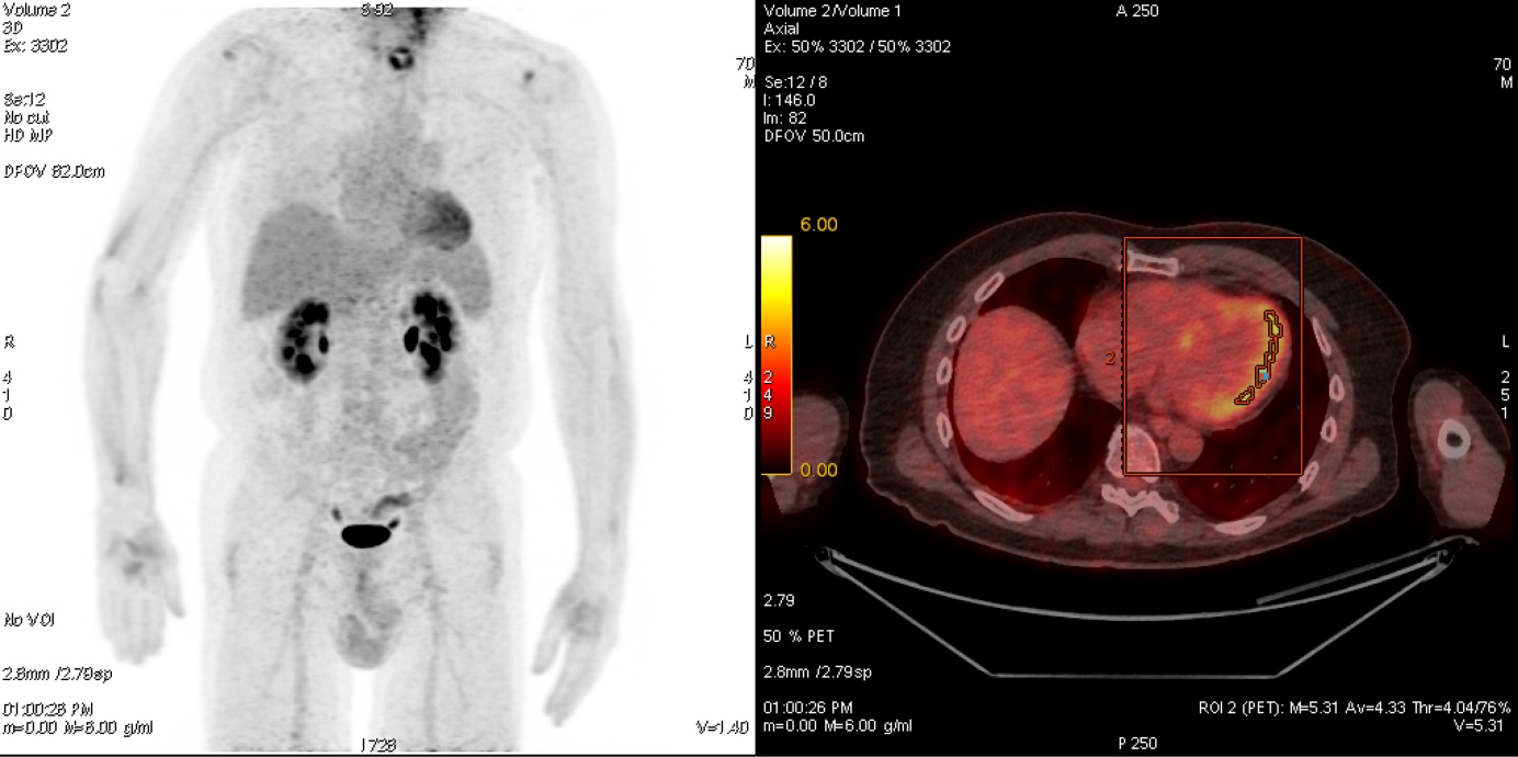
70-year-old patient with Vd.a. cardiac sarcoidosis, which could be clearly diagnosed using 18F-FDG PET/CT: The maximum intensity projection (left) shows clear metabolic activity in the left ventricular myocardium. In the fused PET/CT (right), the increased activity (yellow) is mainly seen in the lateral wall of the myocardium. The activity can be quantified simultaneously using PET (SUVmax here 5.31) and is therefore extremely helpful for therapy monitoring in particular.

In selected cases, particularly in the case of myocarditis or sarcoidosis, it may be useful to combine PET and cardiac MR, which is easy to perform in the combined PET/MR device used at our clinic in the Wagi-Areal in Schlieren, increases the diagnostic accuracy compared to the individual examinations and expands the range of questions that can be answered overall.

63-year-old patient with post-biological aortic valve replacement. Echocardiographic evidence of a possible paravalvular abscess, which can be clearly diagnosed by 18F-FDG PET/CT. Maximum intensity projection (MIP) with clear focal metabolic activity in projection onto the aortic root (A), which can be clearly localized in the series fused with the CT (B).

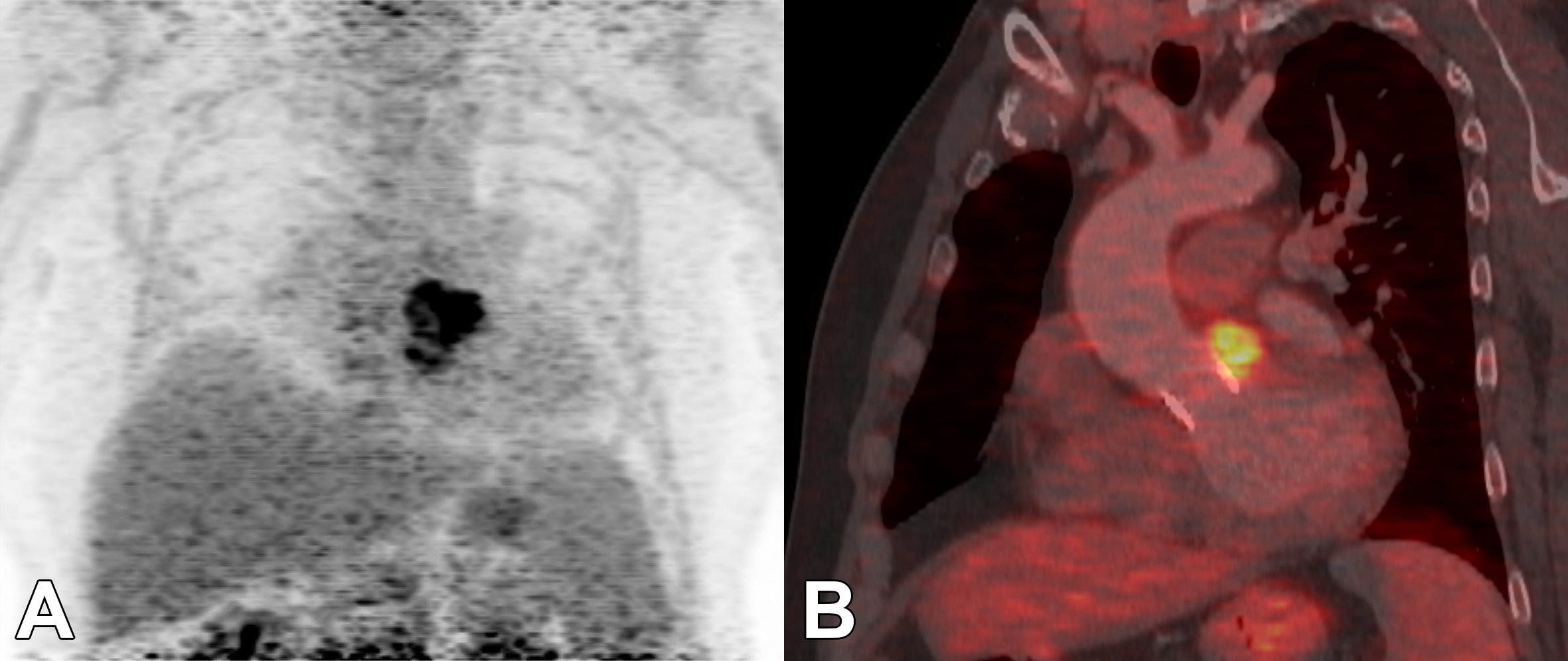
63-year-old patient with st.n. biological aortic valve replacement. Echocardiographic Vd.a. possible paravalvular abscess, which can be clearly diagnosed using 18F-FDG PET/CT. Maximum Intensity Projection (MIP) with clear focal metabolic activity in projection onto the aortic root (A), which can be clearly localized in the series fused with CT (B).

Responsible specialist

Philipp Kaufmann, Prof. Dr. med.

Director of Department, Department of Nuclear Medicine
Head of Outpatient Medicine

Tel. +41 44 255 41 96
Specialties: Specialist for non-invasive cardiac imaging (PET, SPECT, CT, MRI), Specialist FMH for Nuclear Medicine, Cardiology and Internal Medicine

Ronny Ralf Büchel, Prof. Dr. med.

Senior Attending Physician, Vice Director of Department, Department of Nuclear Medicine

Tel. +41 43 253 87 89
Specialties: Specialist in multimodal non-invasive cardiac imaging (PET, SPECT, CT, MRI)., FMH specialist in nuclear medicine and cardiology., Research group: Hybrid and molecular cardiac imaging

Aju Pazhenkottil, PD Dr. med.

Attending Physician with extended responsibilites, Department of Nuclear Medicine
Attending Physician, Department of Consultation-Liaison Psychiatry and Psychosomatic Medicine

Tel. +41 44 255 39 50
Specialties: Cardiac imaging (cardiac CT/NUK/MRI), Echocardiography, Psychocardiology

Andreas Giannopoulos, Dr. med. Dr. med. sc.(GR)

Attending Physician, Department of Nuclear Medicine

Tel. +41 43 253 08 43
Specialties: Specialist in multimodal non-invasive cardiac imaging (PET, SPECT, CT, echocardiography), Specialist in cardiology, Research group: Hybrid and molecular cardiac imaging

Dominik Benz, PD Dr. med.

Attending Physician, Department of Cardiology
Attending Physician, Department of Nuclear Medicine

Tel. +41 43 253 11 91
Specialties: Echocardiography, Cardiac imaging (MRI, CT, NUK), Cardiac amyloidosis

For patients

As a patient, you cannot register directly for a consultation. Please get a referral from your primary care physician, specialist.

Tel. +41 44 255 35 55
Contact form

For referrering physicians

Simply assign your patient via registration form.

Tel. +41 44 255 35 55

Medical information hotline: 08.00-18.00 hrs: +41 44 255 15 01

Registration form

Responsible Department

Related diseases