However, ATTR amyloidosis in particular can be easily diagnosed by means of a simple skeletal scintigraphy. Although the mechanism by which the bone-specific tracers (e.g. DPD) attach to the amyloid in the heart is still unclear, skeletal scintigraphy shows a positive predictive value of 100% (with simultaneous exclusion of AL amyloidosis, i.e. negative serum and urine electrophoresis; Gillmore et al. Circulation. 2016 Jun 14;133(24):2404-12) for the diagnosis of ATTR amyloidosis (Figure 5). No special preparations are required for this.
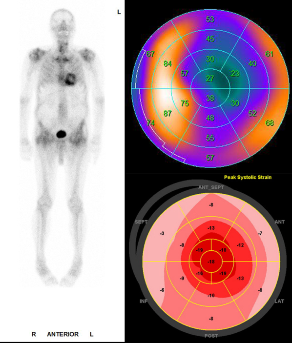
74-year-old patient with hypertrophic left ventricular myocardium. The differential diagnosis was hypertensive heart disease with known hypertension, but echocardiography was suspicious for amyloidosis. The whole-body skeletal scintigraphy (left) clearly shows accumulation of the bone tracer in the myocardium. The local accumulation in the left ventricular myocardium (shown as a so-called polar plot, top right) is mainly septal and lateral, whereas the apex remains unaffected. The scintigraphically detected enrichment corresponds strikingly well with the echocardiographic strain map (bottom right). As there was no evidence of AL amyloidosis in the electrophoresis, the diagnosis of cardiac ATTR amyloidosis could be clearly established.