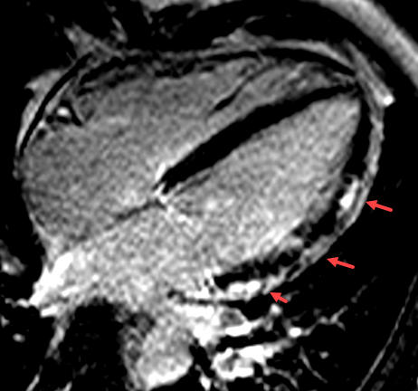
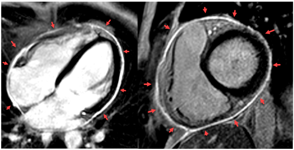
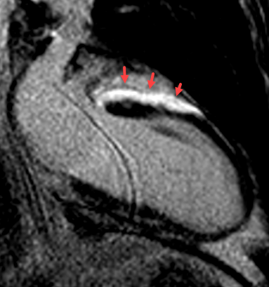
One of the most common causes of inflammation of the heart muscle (myocarditis, Figure 1) or the pericardium (pericarditis, Figure 2) is infection by viruses or bacteria. Autoimmune diseases or toxic reactions are less frequently responsible. Sarcoidosis(Figure 3) as a granulomatous multisystem disease poses a particular diagnostic challenge. Imaging can be used to assess various organ involvement, in particular cardiac manifestations and their prognostic implications. Other important diseases are endomyocardial fibrosis or changes in the heart, e.g. in the case of a COVID-19 infection.
Cardiac magnetic resonance imaging (MRI) is the gold standard for the imaging diagnosis of these diseases.
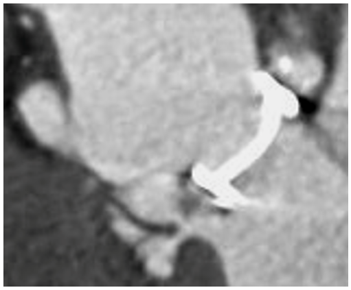
In contrast, cardiac CT is used for imaging inflammations or infections of the heart valves (so-called endocarditis). With CT it is possible to detect changes such as vegetations, pseudoaneurysms or valve dehiscences(Figure 4). This is also possible with CT in patients who have already had a heart valve replaced or operated on.


