Diagnostics and specialized therapy control for liver tumors

The spectrum of liver tumors ranges from completely harmless to malignant tumors that require treatment. Accordingly, the most accurate diagnosis possible is essential.

Diagnostics

In liver diagnostics, ultrasound is used as a so-called “screening” examination. With high-resolution sonography devices and additional contrast enhancement using a contrast agent specially developed for ultrasound, small tumors can increasingly be detected. In many cases, magnetic resonance imaging (MRI), which, like ultrasound, does not require any radiation exposure, is necessary for the precise characterization of the tumour. The so-called “liver-specific” (“hepato-biliary”) contrast agent is used for the examination of liver lesions in MRI. This “liver-specific” contrast agent is absorbed by healthy liver cells 10-20 minutes after intravenous administration, while tumor cells in malignant tumors do not absorb this contrast agent or only to a small extent. With the help of this specific contrast medium information and the excellent organ contrast in the MRI, normal liver tissue or benign liver tumors can be differentiated from malignant liver tumors with a high degree of diagnostic accuracy.

In addition to the characterization of liver tumors, MRI also provides comprehensive information for planning liver surgery, such as the exact number of liver tumors, the positional relationship and exact course of the hepatic arteries, portal veins, hepatic veins and bile ducts, as well as the liver volume.
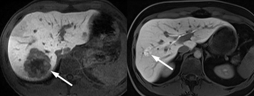
Malignant liver tumor (liver metastasis) with slight accumulation of the “liver-specific” contrast agent (left). Benign liver tumor (focal nodular hyperplasia) with strong accumulation of the “liver-specific” contrast agent (right).
Specialized therapy control

Over the last few years, targeted local therapy procedures in interventional radiology have been used more and more frequently in the treatment of liver tumors. In these therapeutic procedures, such as chemoembolization, radioembolization or radiofrequency ablation, the blood supply to the tumour is specifically interrupted or the tumour tissue is killed. Our and international studies have shown that specialized, so-called functional CT or MRI are advantageous for monitoring the success of these procedures, and that a successful therapy can thus be identified at an early stage.

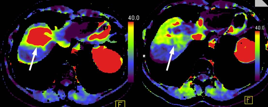
Using functional CT perfusion, the blood flow to the liver tumor can be precisely measured and displayed in a color-coded perfusion image. If the liver tumor responds to local therapy, CT perfusion reveals a significant reduction in tumor perfusion at an early stage.

CT perfusion of a malignant liver tumor before treatment (left) with strong tumor perfusion and four weeks after treatment by radioembolization (right) with good therapy response and significant reduction of tumor perfusion.

Responsible squad

Andreas Hötker, PD Dr. med.

Senior Physician, Institute for Diagnostic and Interventional Radiology

Tel. +41 43 253 85 12
Specialties: Urogenital radiology, Gastrointestinal radiology, Magnetic resonance imaging

Soleen Stocker, PD Dr. med.

Senior Attending Physician, Institute for Diagnostic and Interventional Radiology

Tel. +41 44 253 11 67
Specialties: Urogenital and gynecological imaging, Abdominal imaging, Oncological imaging

Daniel Stocker, PD Dr. med.

Senior Attending Physician, Institute for Diagnostic and Interventional Radiology

Tel. +41 43 253 25 99

For patients

As a patient, you cannot register directly for a consultation. Please get a referral from your primary care physician, specialist.

For referrering physicians

University Hospital Zurich
Institute for Diagnostic and Interventional Radiology
Raemistrasse 100
8091 Zurich

Tel. +41 43 254 41 10
Patient registration form

Related diseases