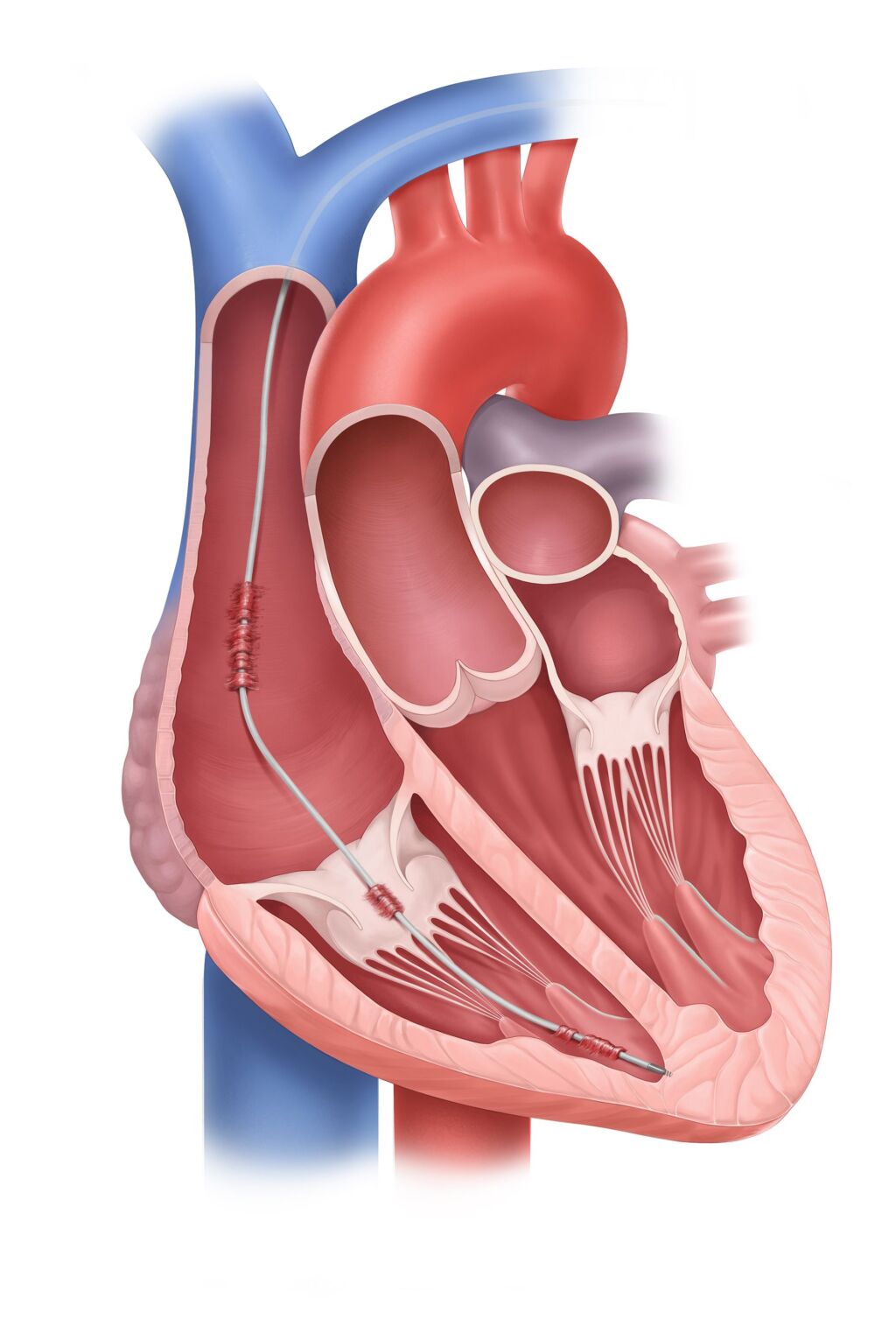
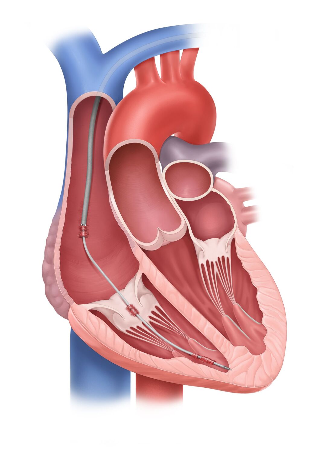
The weak point of pacemaker and defibrillator devices are the so-called electrodes (probes), which lead from the battery of the device via the veins into the heart. Infections may occur or the electrodes may become dysfunctional and no longer work. In such situations it may be necessary to remove the affected electrodes. However, as these grow together with the body tissue over the years, trained medical professionals and special material are required to perform such electrode extractions. The aim of this intervention is to gently and safely remove all the electrodes from the heart via the veins. This is usually successful in 98% of cases with a very low complication rate.
Preparation
A consultation appointment is organized a few weeks before the procedure to discuss the procedure with the medical staff and clarify any unanswered questions. In addition, the necessary preliminary examinations are carried out, which include a chest X-ray and a heart ultrasound examination called echocardiography. Echocardiography is used to check the current condition of the heart, including the pumping capacity and the possible influence of the electrodes in the heart on the function of the heart valves. We will then discuss in detail how the procedure will proceed and what complications can potentially be expected. There is also a discussion about which new device should be implanted again.
Procedure
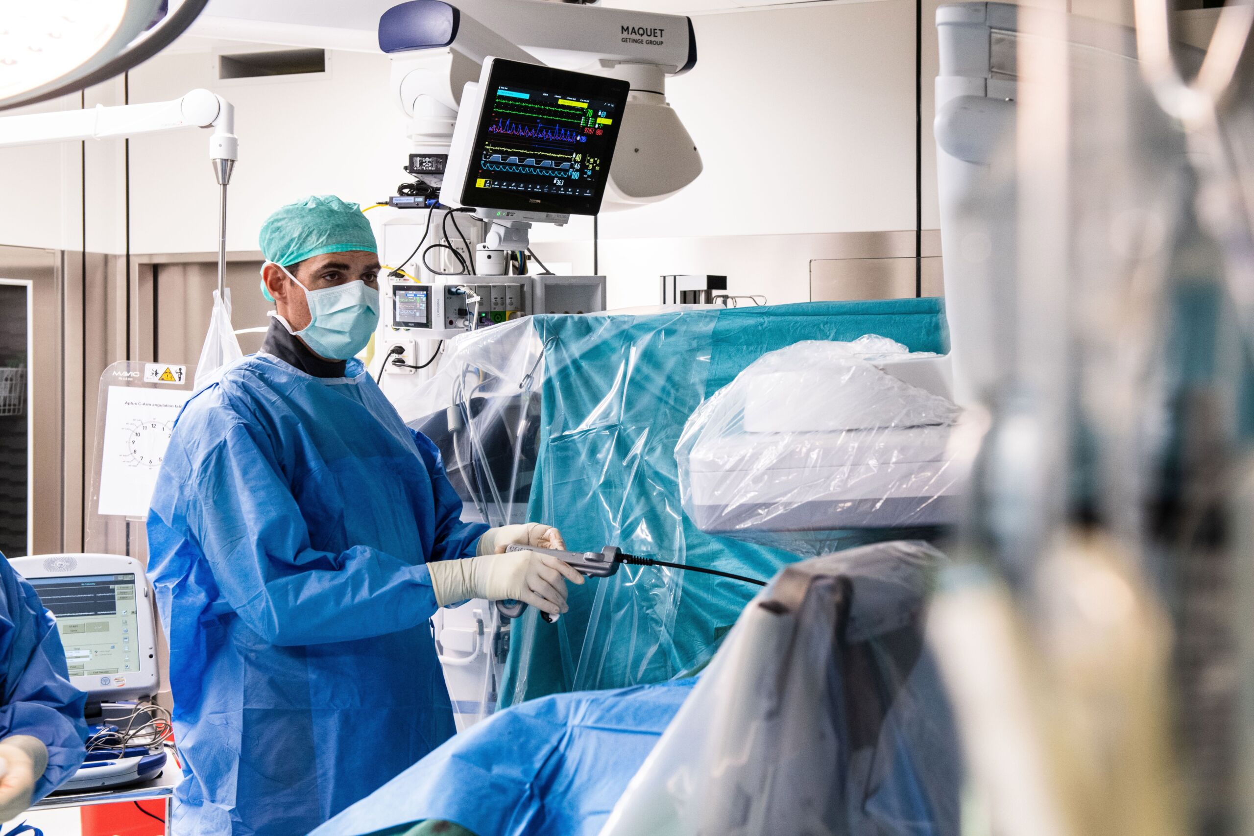
The procedure is carried out in a special operating theater using state-of-the-art X-ray diagnostics. Thanks to the support of the cardiac anesthesia, cardiac surgery and cardiotechnical teams, these operations can be performed under the best cardiovascular monitoring. Cardiac anesthesia takes care of the anesthesia during the procedure and monitors the patient throughout the intervention. For this purpose, a swallowing ultrasound (transesophageal echocardiography) is used as standard to continuously monitor the functionality of the heart and detect possible complications at an early stage. The procedure begins with the electrodes being prepared for extraction. After a skin incision and opening the pocket where the device is located, the electrodes are removed from the battery. A thin wire is then inserted through the probes to stabilize the electrodes. A special extraction tool can now be inserted via the probes and guided forward step by step to the end of the electrodes in the heart. This allows the electrodes to be removed from the body and, if necessary, a new device to be implanted.
After the procedure
After the operation, patients usually wake up in the operating room and are then transferred to the cardiology monitoring ward, where the medical staff take care of the postoperative course until the next morning before the patients are transferred to the normal ward. Another chest X-ray is taken to check the position of the new implant and the device is checked. Depending on the clinical situation, further examinations may be necessary. In stable situations, the patient can then be transferred back to the normal ward the next day and leave the hospital the following day.
National and international collaboration
These highly specialized procedures are only performed in a few centers in Switzerland and the USZ is one of the leading institutions in this field. An intensive exchange with other specialists is essential. In order to guarantee quality and promote exchange, a nationwide collaboration has been established in recent years together with other service providers in Switzerland. In addition, there is an intensive international exchange with, among others, the cardiac surgery department at the renowned Charité Clinic in Berlin, one of the world’s largest electrode extraction centers, and our experts are regularly invited to give presentations and participate in discussions at national and international congresses.


