What is esophageal cancer?
Esophageal cancer (esophageal carcinoma) is a malignant tumor of the esophageal mucosa (esophagus). Experts distinguish between two types of esophageal cancer:
- Adenocarcinomas: These develop from glandular cells. They occur in the lower section of the esophagus.
- Squamous cell carcinomas: Squamous cell carcinomas develop in the upper and middle esophagus
The most important risk factors for squamous cell carcinoma of the oesophagus are frequent consumption of high-proof alcohol and smoking. Risk factors for adenocarcinoma are chronic heartburn and obesity. Esophageal cancer gradually narrows the esophagus. As the oesophagus is a very flexible organ, those affected usually only have problems absorbing food at a very advanced stage.
Frequency of esophageal cancer
In Switzerland, around 570 people are diagnosed with esophageal cancer every year. This corresponds to around one percent of all cancer cases in Switzerland. Three quarters of men are affected, one quarter of women. Esophageal carcinomas predominantly occur at an older age.
Esophageal cancer: causes and risk factors
The causes of esophageal cancer are not yet fully understood. The risk factors include:
- Alcohol consumption,
- Smoking,
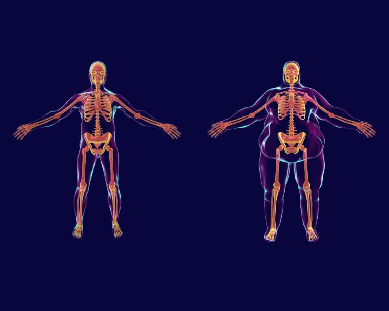
- Overweight,
- very hot drinks,
- chronic heartburn,
- Partial closure of the entrance to the stomach,
- Acid or alkali burns,
- Tumors in the mouth and neck area,
- Radiation in the neck and chest area,
- Barrett’s syndrome (abnormally altered mucous membrane of the lower esophagus),
- Congenital malformation of the esophagus (so-called achalasia) or acquired changes (e.g. scars).
Gastric and esophageal tumor center
At the USZ, numerous specialist departments have joined forces to form a gastric and esophageal tumor center. The center is certified according to the guidelines of the German Cancer Society (DKG). A team of experts specializing in the medical treatment of oesophageal cancer works closely together here for the benefit of our patients. At DKG-certified centers, patients are treated according to strict quality criteria and, according to current studies, have a better chance of survival on average.
Esophageal cancer often without symptoms at first
In many patients, esophageal cancer initially causes hardly any symptoms. This is why a disease is only noticed when it is already advanced.
Typical warning signs of esophageal cancer are:
- Difficulty swallowing and pain when eating due to narrowing of the esophagus,
- frequent ingestion,
- Loss of appetite and weight loss,
- Hoarseness,
- vomiting for no reason,
- Blood in the stool (tarry stool),
- A feeling of pressure or pain behind the breastbone and in the back when the tumor constricts the esophagus and food accumulates in the esophagus; these symptoms are usually less severe with liquid or soft foods such as soups or porridge.
Please do not hesitate to visit us if you have complaints over a longer period of time.
Esophageal cancer: Diagnosis with us
To diagnose esophageal cancer, we will first ask you about your symptoms. Various examinations are also possible.
Esophageal cancer: prevention, early detection, prognosis
Smoking and excessive alcohol consumption increase the risk of esophageal cancer. You can prevent esophageal cancer by avoiding these addictive substances. Chronic heartburn also increases the risk of illness. If stomach acid repeatedly flows into the esophagus (so-called reflux), the acid burns the mucous membrane. This can develop into beret syndrome. In Barrett’s syndrome, the esophageal mucosa may change into a precursor of esophageal cancer. Consult us if you suffer from heartburn over a longer period of time.
Progression and prognosis of esophageal cancer
If esophageal cancer (esophageal carcinoma) is detected in time, the chances of recovery are good. But this is rarely the case. The more advanced the esophageal cancer, the more difficult it is to treat.
Second opinion for esophageal cancer
When a cancer diagnosis is made, a second medical opinion is an important decision-making tool. The Comprehensive Cancer Center Zurich supports you with a professional expert opinion. They receive a thorough analysis of the situation as well as personal advice and quick answers to their questions.
Esophageal cancer: Treatment
Local treatment of esophageal cancer is usually successful. Provided that no metastases have formed in other organs and the original tumor has not spread too far. Which therapy is suitable for patients depends on the type of tumor and the stage of the tumor.
