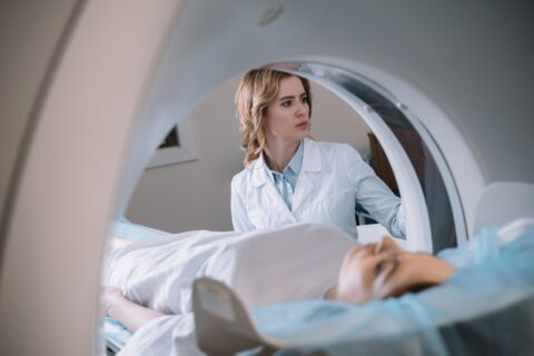
MRI of the pelvic floor can be used to examine the pelvic floor muscles and the pelvic organs. On the one hand, the quality of the pelvic floor muscles can be visualized, and on the other hand, the mobility and tightness of the pelvic floor muscles can be examined using dynamic MRI images. MR defecography is used to examine the emptying of the rectum. This can reveal coordination disorders of the muscles involved and other causes of bowel evacuation disorders. The dynamic MRI images can also be used to assess prolapse of the pelvic floor and pelvic organs (e.g. the uterus).
The examination is performed in a conventional MRI machine in a supine position with slightly bent legs and takes about 25 minutes. After taking pictures at rest, instructions are given via a microphone to tense the pelvic floor, press and finally empty the rectum. The movement of the pelvic floor and the organs is recorded with the MRI device.
