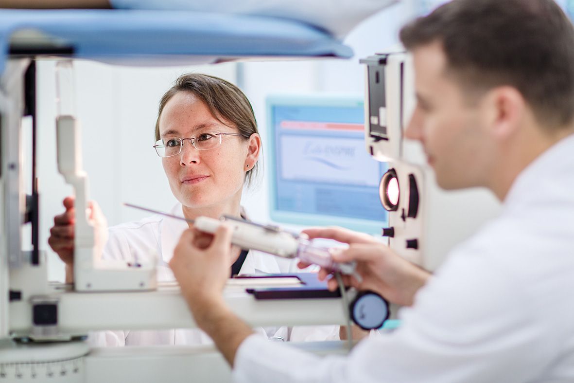
Ultrasound-guided vacuum biopsy
During a vacuum-assisted biopsy, the patient lies on her back and the breast findings are visualized with the ultrasound device. After disinfection and local anesthesia, the needle is inserted under the lesion and removed step by step under ultrasound visual control.

Stereotactic vacuum biopsy
Findings that are only visible on mammography, such as microcalcifications, can be clarified using stereotactic vacuum biopsy.
During the stereotactic vacuum-assisted biopsy, the patient lies on her stomach and the breast findings are visualized with the mammography device, which is positioned underneath the table. After disinfection and local anesthesia, the needle is guided to the findings after calculating the setting with the computer and samples are taken from this area. The samples are then x-rayed in a separate unit to ensure that the findings, e.g. microcalcifications, are contained in the sample.
